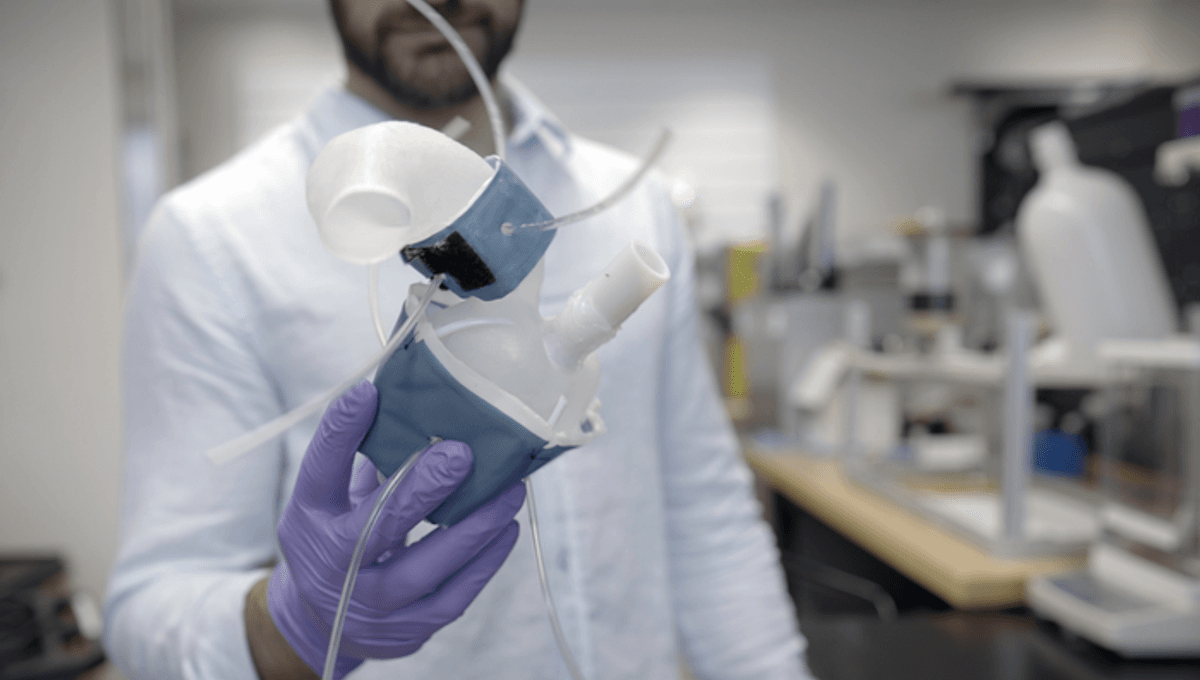
Valentine’s Day may have passed, but it looks like engineers at MIT have found another way for you to give your heart to a loved one in the future – 3D-print it!
Every heart is unique. Not only does the organ’s overall shape and size vary from person to person and change with age, but even their texture is different depending on if you are male or female. This level of idiosyncrasy is normal – but in some cases, particularly for people living with heart disease or congenital defects, it can lead to extra strain as their hearts and associated blood vessels have to work harder to overcome any limits to their function.
Now, engineers led by Ellen Roche, a mechanical engineering professor at MIT, have developed a way to custom print robotic hearts that can be used to replicate a patient’s heartbeat and the way it would naturally pump blood around the body.
The procedure involves converting scanned images of a patient’s heart into a three-dimensional computer model that can then be printed using a rigid photopolymer, a type of polymer that solidifies under ultraviolet light.
Once printed and cured, the photopolymer is extremely pliable. When used to create the modeled hearts, it results in a soft, flexible shell that is a perfect reproduction of the patient’s own heart. This same method can be used to print a patient’s aorta, the artery that carries blood away from the heart to the rest of the body.
The team can then attach fabricated sleeves, similar to blood pressure cuffs, around the new heart and aorta. These sleeves have a precisely patterned bubble wrap-like underside which, when connected to a small air pump system, rhythmically inflate causing the replica heart to constrict and contract, much like its natural pumping action.
“All hearts are different,” Luca Rosalia, a graduate student in the MIT-Harvard Program in Health Sciences and Technology said in a statement. “There are massive variations, especially when patients are sick. The advantage of our system is that we can recreate not just the form of a patient’s heart, but also its function in both physiology and disease.”
The researchers paid particular attention to aortic stenosis, a condition where the aortic valve is narrowed and does not open fully. This reduces or blocks blood flow from the heart to the aorta where it cannot flow to the rest of the body. This can be caused by aortic valve fibrosis and calcification, and can present patients with increased fatigue, shortness of breath, angina, syncope, and even heart failure. Aortic stenosis is often treated by implanting synthetic valves designed to widen the aorta.
Using the 3D printer method, the team of engineers was able to inflate a separate sleeve wrapped around a printed aorta and inflated it in a way that imitated aortic stenosis. Professor Roche and her colleagues then implanted synthetic valves into the printed aorta modeled on specific patients suffering from the condition. They found that, when they activated the pump action, the implants behaved the same way as they would in an actual patient. The team was also able to use an actuated printed heart to compare implant sizes to see which options performed best for the patient.
It is hoped that this innovation will allow them to collaborate with doctors to find the best implants for their specific hearts.
“Patients would get their imaging done, which they do anyway, and we would use that to make this system, ideally within the day,” says Christopher Nyugen, co-author of the team’s recent publication. “Once it’s up and running, clinicians could test different valve types and sizes and see which works best, then use that to implant.”
The paper has been published in the journal Science Robotics.
Source Link: 3D-Printed Hearts Are The Future Of Valve Replacement Surgery