


Do you have an eye for the wonders of the miniature world? Are you able to capture the beauty of things too small for human perception? Then now is the time to showcase your talent. Grab your microscopes and camera and join Nikon as they celebrate 50 years of their Small World Competition. This time, you could be the big winner in the world of tiny wonders.
Small worlds are a big deal
The Small World Competition started in 1975 to recognize and applaud the skill of photography performed through the light microscope. Since then, this competition has become a place to exhibit the staggering beauty of this unseen world in a way that blends art with science.
Each year, participants from a broad range of scientific disciplines submit an ever more diverse range of images – from the intricate patterns of a rodent optic nerve to the delicate but vibrant details of a live oak leaf, or the mesmerizing crystalline structure of a frozen water droplet – to show that even the smallest subjects can leave big impressions.
Since 2011, Nikon has also run the Small World in Motion competition, which showcases movies or digital time-lapse photography taken through the microscope. These submissions add a distinct visual dimension to otherwise invisible activities taking place around or inside us.
50 years of mesmerizing microscopy
Nikon’s Small World is the oldest and largest photomicrography competition in the world. As such, it is not only a catalog of luminous and exquisite images of rarely seen specimens, but it is also a historical record of the subtle advancements in technology and scientific precision.
“Nikon’s Small World Competition beautifully illustrates how far microscopic imaging has come in the past 50 years,” Tom Hale, Senior Journalist at IFLScience and judge for the 2019 Small World Competition, explained. “Looking at the hundreds of stunning images that have been entered over the decades, you get a sense of how science, technology, and art are deeply intertwined with each other.”
“Over the 50-year course of this competition,” added Eric Flem, Communications and CRM Manager, Nikon Instruments, “the gallery has mirrored not only the advances in technology in microscopy imaging, but has also acted as a barometer for various scientific disciplines that have been opened up as microscope technologies made imaging in these areas possible.”
The developments in microscopes and digital cameras have transformed the field from a highly specialized practice to something that is more accessible to wider groups of people. This does not mean that photomicrography is easy, mind you. It still demands skill and knowledge to perform well, but digital cameras allow scientists, artists, and anyone else to view and review results faster than before.
This has also meant a change in the subjects being photographed. As Flem explained to IFLScience: “As the competition evolved through the years there has been a shift to more biological samples as technology has made the capture of these types of images easier and more widely available to scientists, artists, and hobbyists.”
Just a casual glance at the winning entries for each year is enough to illustrate this. In 1975, the winning image was submitted by James Dvorak who presented a mosaic-like image of oxalic acid crystals during precipitation. Dvorak captured this image by transmitting polarized light through a quartz wedge. Then, 10 years later, Jonathan Eisenback presented a formalin-fixed whole mount of a spiral nematode that he captured using multiple exposures. Throughout these years, the majority of images were of static structures, but you can see the complexity and clarity of the images developing over this time. Today, the situation is much different. The images submitted are crisp and sharp compared to earlier entries, and they are produced using a wider range of techniques.
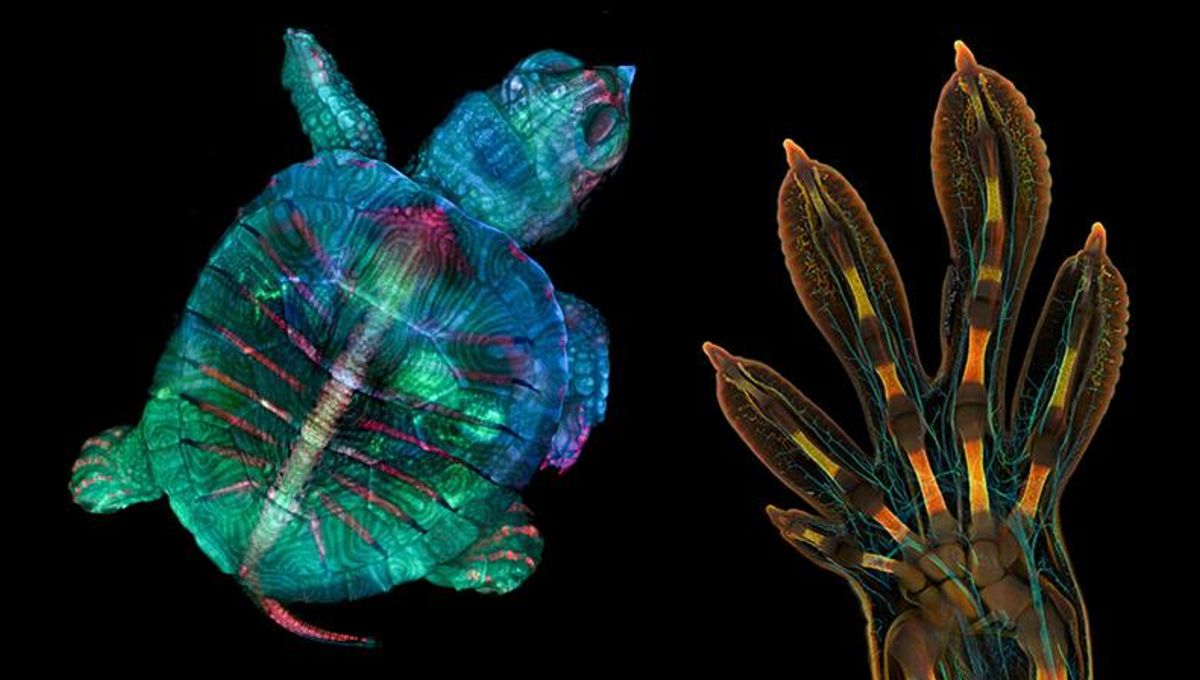
For instance, the winner of the 2022 Small World Competition used high-resolution microscopy and image-stitching to capture the embryonic hand of a Madagascar giant day gecko. To achieve this, Grigorii Timin, supervised by Michel Milinkovitch, performed whole-mounted fluorescent staining and tissue clearing to capture the hand with a confocal microscope.
“The scan consists of 300 tiles, each containing about 250 optical sections, resulting in more than two days of acquisition and approximately 200 GB of data”, Timin told Nikon.
How to enter
Entries for this year’s competition, both Small World and Small World in Motion, are open until April 30, 2024. Anyone over the age of 18 with an interest in microscopy and photography can take part, and all participants receive a calendar for entering. For more information about prizes or any other questions, visit Nikon’s Small World FAQ page.
Competitors are welcome to use any type of light microscope and technique, including phase contrast, polarized light, fluorescence, interference contrast, darkfield, confocal, deconvolution, and mixed techniques. Electron microscope and macro images are not eligible.
So, grab your favorite specimens and take part in the ultimate showcase of all things miniature and marvelous. Remember, entry is open until April 30, 2024.
This article includes sponsored material. Read our transparency policy for more information.


Source Link: Celebrating 50 Years Of Microscopic Masterpieces: Nikon’s Small World Competition Is Open For Submissions