In a world first, scientists have successfully grown human brain organoids – so-called “mini brains” – from human fetal tissue. The organoids are only about the size of a grain of rice, but they have the potential to offer a whole new way of studying brain development and disease.
Organoid research has exploded in recent years. From stomachs, to kidneys, to a whole “body-on-a-chip”, being able to generate miniature replicas of organs has the potential to revolutionize medical research.
Brain organoids that start from human stem cells have previously been shown to respond to visual stimuli; used to repair injured rat brain tissue; and been infected with COVID-19 to study the damage it can do to the nervous system. A mini brain was even fused with computer hardware to create a hybrid biocomputer.
Despite these incredible advances, however, there’s been a limitation to how human brain organoids can be grown. Up to now, the only option has been to use embryonic or pluripotent stem cells that are prodded down the correct developmental pathway using a cocktail of very specific molecules, which can take a long time to determine.
Now, scientists have found a way to produce mini brains directly from fetal brain tissue.
“Until now, we were able to derive organoids from most human organs, but not from the brain – it’s really exciting that we’ve now been able to jump that hurdle as well,” explained project co-lead Professor Dr Hans Clevers in a statement.
The team, from the Princess Máxima Center for Pediatric Oncology in the Netherlands, figured out that the key was to use small pieces of whole tissue, rather than individual cells as is required for organoids created from other organs. Under the correct growth conditions, these tissue fragments self-organized into complex 3D brain structures.

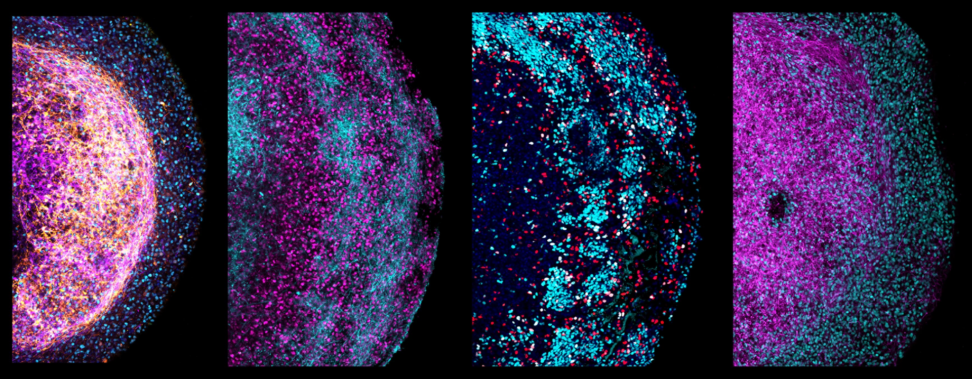
The complexity of one of the organoids can clearly be seen in these different sections.
Although tiny, the organoids contained multiple different cell types, and retained specific characteristics of the parts of the brain from which they were derived. For example, they still responded to various signaling molecules that direct brain development as a fetus grows – this means they could be used to provide new insights into this highly complicated process.
Since the organoids are quick to grow, the team decided to test their potential in modeling brain cancer. Using CRISPR-Cas9 gene editing, they mutated a cancer gene called TP53. After three months, the mutated cells had taken over, just as cancer cells do.
Later, they used the same technique to alter three genes associated with glioblastoma, a type of brain cancer, and tested the effect of some cancer drugs on the mutant organoids. This is just one other way that these mini brains could be used in scientific research in the future.
The organoids will happily grow in the lab for more than six months and can be multiplied, meaning that scientists can repeat their experiments on similar organoids to increase the reliability of their results. The tissue used to grow the organoids in the first place is not an infinite resource, so it’s important that its use is maximized as much as possible.

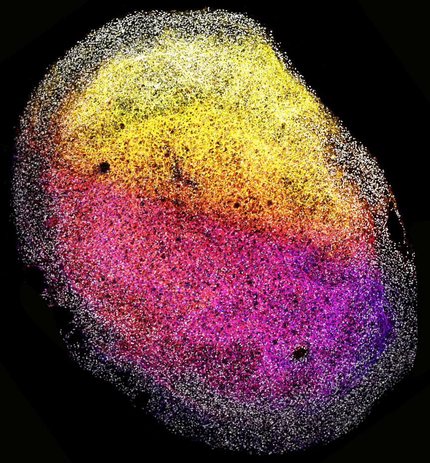
A whole organoid.
The fetal tissue used in the research was donated by people undergoing pregnancy terminations between 12 and 15 weeks of gestation. The donors were kept totally anonymous and had given full consent to the use of the tissues in organ development research.
The team hope to continue exploring the potential of their mini brains. They have also been working with bioethicists and aim to continue this collaboration to shape the future of research in this area.
“Being able to keep growing and using the brain organoids from fetal tissue also means that we can learn as much as possible from such precious material,” said co-study lead Dr Delilah Hendriks. “We’re excited to explore the use of these novel tissue organoids for new discoveries about the human brain.”
The study is published in the journal Cell.
Source Link: For First Time, "Mini Brains" Have Been Grown From Human Fetal Brain Tissue