


“Mini brains” composed of lab-grown human cells can integrate into injured rat brains and respond to visual stimuli, a new study has reported. Previous research has shown that integration into developing brains was possible, but this new study is the first to demonstrate the use of this approach to repair a brain injury.
Human brain organoids are miniature versions of brain tissue produced from pluripotent stem cells cultivated in the lab. They might be significantly smaller than the real thing, but organoids do reproduce at least some of the features of natural brain tissue. Some of the different cell types that we would expect to find in the brain, like astrocytes and oligodendrocytes, do appear as the organoids develop; and the structure of the tissues is similar, though less complex than a real brain would be.
In recent years, studies have gradually added to the picture of how these organoids might one day play a part in the treatment of neurological disease. Organoids exhibit neural activity of their own, even when still in the culture dish. They’ve been successfully transplanted into the brains of rodents, and are able to mesh with the surrounding tissue – a recent study even showed that they react to light in the same way as normal neurons.
However, something science had not yet explored was whether organoids would be able to integrate with injured brain tissue. A new study, led by researchers at the University of Pennsylvania, has begun to answer that question.
“We focused on not just transplanting individual cells, but actually transplanting tissue,” said senior author H. Isaac Chen in a statement.
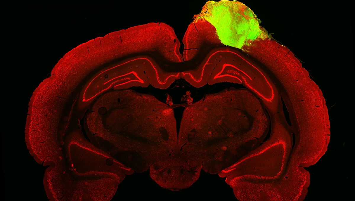
The organoids were grown in the lab for around 80 days before being grafted into their new hosts. The rats used in this experiment were adults, and had sustained injuries to the visual cortex. After three months, the team used a nifty fluorescent-tagged virus, injected into the eye, to track the new connections that the organoids had made with the surrounding tissue.
“By injecting one of these viral tracers into the eye of the animal, we were able to trace the neuronal connections downstream from the retina,” said Chen. “The tracer got all the way to the organoid.”
The big test was to see if the organoid’s neurons could respond to visual stimuli – if so, it would mean that organoids would have the potential to take on the functions of injured brain tissue, and thereby help to repair it. Electrodes were used to probe individual neurons within the organoid, measuring their activity while the animals were exposed to flashing lights and alternating black and white bars.
“We saw that a good number of neurons within the organoid responded to specific orientations of light, which gives us evidence that these organoid neurons were able to not just integrate with the visual system, but they were able to adopt very specific functions of the visual cortex,” Chen explained.
It was a surprising result for the team. “We were not expecting to see this degree of functional integration so early. There have been other studies looking at transplantation of individual cells that show that even nine or 10 months after you transplant human neurons into a rodent, they’re still not completely mature,” Chen said.
Chen hopes that these results will pave the way for further tests of this approach in other brain areas: “Now, we want to understand how organoids could be used in other areas of the cortex, not just the visual cortex, and we want to understand the rules that guide how organoid neurons integrate with the brain so that we can better control that process and make it happen faster.”
The study is published in Cell Stem Cell.


Source Link: Human "Mini Brains" Merge With Injured Rat Brain Tissue And Respond To Light