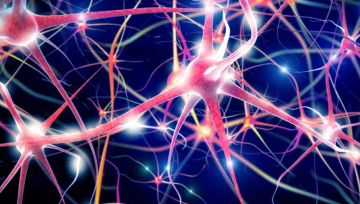
For the first time, researchers have demonstrated that human brain organoids are able to form functional connections with mouse brain tissue and respond to visual stimuli. After implanting the human “mini brains” into the animals’ cortices, the study authors used a variety of imaging techniques to confirm the formation of “human-mouse synapses.”
In recent years, human brain organoids have emerged as a promising model for the study of neurological development and disorders. Created from stem cells, these miniature cortical replicas produce their own neural activity, but had never previously been observed linking up with surrounding tissues to participate in a synchronized response to external stimuli.
The main barrier to demonstrating this phenomenon has always been a technological one, as existing electrode arrays are not subtle enough to record such nuanced activity. However, the researchers were able to overcome this hurdle by using platinum nanoparticles to create transparent graphene microelectrode arrays.
When mice were presented with a white light visual stimulus, the researchers were able to use these implanted electrodes to measure neural activity in the organoids and the surrounding brain tissue simultaneously. In doing so, they revealed that both reacted to the stimulus in the same way.
“No other study has been able to record optically and electrically at the same time,” explained study author Madison Wilson in a statement. “Our experiments reveal that visual stimuli evoke electrophysiological responses in the organoids, matching the responses from the surrounding cortex.”
Using a highly refined microscopy technique called two-photon imaging, the team were able to show that mouse blood vessels had begun to extend into the human brain organoids, providing them with nutrients and energy. Brainwave activity within the organoids also became synchronized to that of the surrounding tissue, indicating that functional connections had been established between the human and mouse cortical tissues within three weeks of implantation.
“With conventional metal electrodes, we would not have had a clear field of view to examine the organoid graft and proximity to sensory cortex,” write the researchers, adding that “the success of our experiments depended on the engineering of flexible, transparent graphene devices.”
The study authors continued their observations for eleven weeks and showed that the human brain organoids become functionally and morphologically integrated into the mouse cortices.
“We envision that, further along the road, this combination of stem cell and neurorecording technologies will be used for modeling disease under physiological conditions at a level of neuronal circuits, examination of candidate treatments on patient-specific genetic background, and evaluation of organoids’ potential to restore specific lost, degenerated, or damaged brain regions,” they write.
The study is published in the journal Nature Communications.
Source Link: Human “Mini Brains” Observed Responding To Visual Stimuli For The First Time