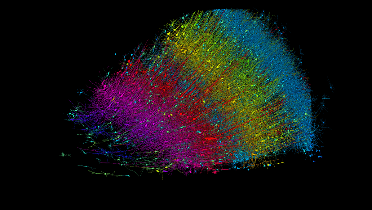
This vivid rainbow of cells represents the largest-ever high-resolution 3D map of a section of the human brain. The largest, yes, but still just a cubic millimeter in size – about half a grain of rice. With this feat, scientists can now see the intricate web of 57,000 cells, connected by 150 million synapses and hundreds of millimeters of blood vessels, that make up this one tiny section of the human cortex.
For the last decade, a collaboration between researchers at Harvard University and Google has been working towards the goal of a complete, high-resolution map of the mouse brain. This is one small but important step on that journey, revealing some of the never-before-seen complexity of a fragment of brain tissue down to the synaptic scale.
“The word ‘fragment’ is ironic,” said senior author Jeff Lichtman, whose team produced the electron microscopy images that form the basis of the new map, in a statement. “A terabyte is, for most people, gigantic, yet a fragment of a human brain – just a miniscule, teeny-weeny little bit of human brain – is still thousands of terabytes.”
With help from AI algorithms developed at Google Research, the imaging from Lichtman’s team at Harvard can be color-coded and reconstructed to reveal unprecedented details.
You might consider that neurons, the archetypal nerve cells, would be the most abundant in the major organ of the central nervous system – the clue is in the name, right? But the team found that these cells were actually outnumbered two to one by the supporting glial cells that help keep the brain’s environment just right. The most plentiful cell type was the oligodendrocytes that produce myelin, the vital insulation around nerve axons.
Among the oddities in the tissue fragment were powerful neurons connected via 50 or more synapses each, swollen axons filled with what the team described in their paper as “unusual material”, and a small number of axons arranged into “extensive whorls”. Since the sample of tissue was originally taken from a patient with epilepsy, it’s unclear whether these features are related to that condition or not.
The importance of mapping down to this level of detail is that it will hopefully provide researchers of the future with new insights into how small-scale connections within brain tissue could potentially be having a big impact, affecting major functions and contributing to disease.
This branch of science is called “connectomics”. Other advances in the field in recent years have seen a massive international project to map the total connections within the human brain – just as we’ve mapped the human genome – and the publication of the first complete map of an insect brain.
Add this to feats like last year’s announcement of a brain cell atlas, and scientists are now able to look more deeply than ever before inside our bundle of “little gray cells”.
To further this aim, and to broaden access to these methods to as many scientists as possible, the Harvard and Google team have developed a series of analytical tools that are publicly available. “Given the enormous investment put into this project, it was important to present the results in a way that anybody else can now go and benefit from them,” said Viren Jain, a member of the Google Research group.
The map of the entire mouse brain that is the end goal of this particular project will generate about 1,000 times the amount of data produced from this 1-cubic-millimeter human brain fragment, so there is still some way to go.
“Without question, approaches to uncovering the meaning of neural circuit connectivity data are in their infancy,” the authors acknowledge, “but this petascale dataset is a start.”
The study is published in the journal Science.
Source Link: Largest-Ever 3D-Mapped Piece Of The Human Brain Could Still Fit On A Grain Of Rice