Microscopes are an iconic instrument in science and since their development over 400 years ago, they have proven to be vital for many key biological discoveries. These optical enhancers have relied heavily on advances in lens technologies and although their history is complex, together they have changed how we see the world.
The first microscopes
As with the telescope, it is unclear who first invented the microscope, but the Dutch spectacle maker Zacharias Janssen and his father Hans are thought to be the individuals who made the earliest compound microscopes at the end of the 16th century. The Janssen’s invention involved using two lenses – once at the top and one at the bottom – in a tube that would magnify an object when looked through. The magnification was relatively low, only between 3x and 9x (which resulted in pretty poor image quality), but the development nevertheless had huge impacts for the future. In particular, the arrangement of the two lenses connected by a tube would remain the standard design for centuries to come.
One of the most substantial issues facing these early magnifying devices was that, although they could enhance the size of an object being observed through them, they could not increase the resolution. Coupled with this, many microscope lenses created aberrations and distortions. This meant the images they produced were blurry and unclear. This remained such a persistent issue that many researchers refused to use them all the way into the 19th century, as they could not trust what they were seeing. Despite this, developments in lens grinding techniques improved microscopes throughout the 17th century, resulting in 200x magnification.
In the late 1660s, these improvements in lenses allowed for new discoveries. In 1665, the English polymath Robert Hooke published the first important book on microscopy, his Micrographia, which contained large, finely detailed illustrations of specimens Hooke had examined under a microscope of his own design. Hooke chose some “interesting” subjects for his observations, which we might think of a pretty gross until we view them in his work. For instance, he looked at the surface of frozen urine, the eye of a grey drone-fly, moss, a woodlouse, an ant, and, famously, a flea. These images are remarkable and capture the wonder that must have been felt for seeing things that are usually far too small for our eyes. Moreover, Hooke brings the images to life with witty and artistic language that demonstrates their splendor.
Aside from conveying the beauty of the very small things he observed, Hooke is also famous for coining the term “cell”, which we still use today in the biological sciences to describe the smallest unit that can live on its own and that is present in all living organisms. For Hooke however, he used “cells” to describe the tiny pockets he saw in the structure of cork, which reminded him of the cells in a monastery.
Following this, in 1676, a Dutch cloth merchant who had taken an interest in science, Antonie van Leeuwenhoek, improved microscopes by designing his own version that relied on a single high-quality lens that functioned more like a magnifying glass. These hand-ground lenses offered an incredible (for their time) 200 times magnification, which allowed Leeuwenhoek to observe various specimens, many of which had never been observed before. These included animal and plant tissues, as well as red blood cells. Significantly, Leeuwenhoek was also the first person to describe and subsequently study bacteria, which had massive implications for the future fields of microbiology and modern medicine.
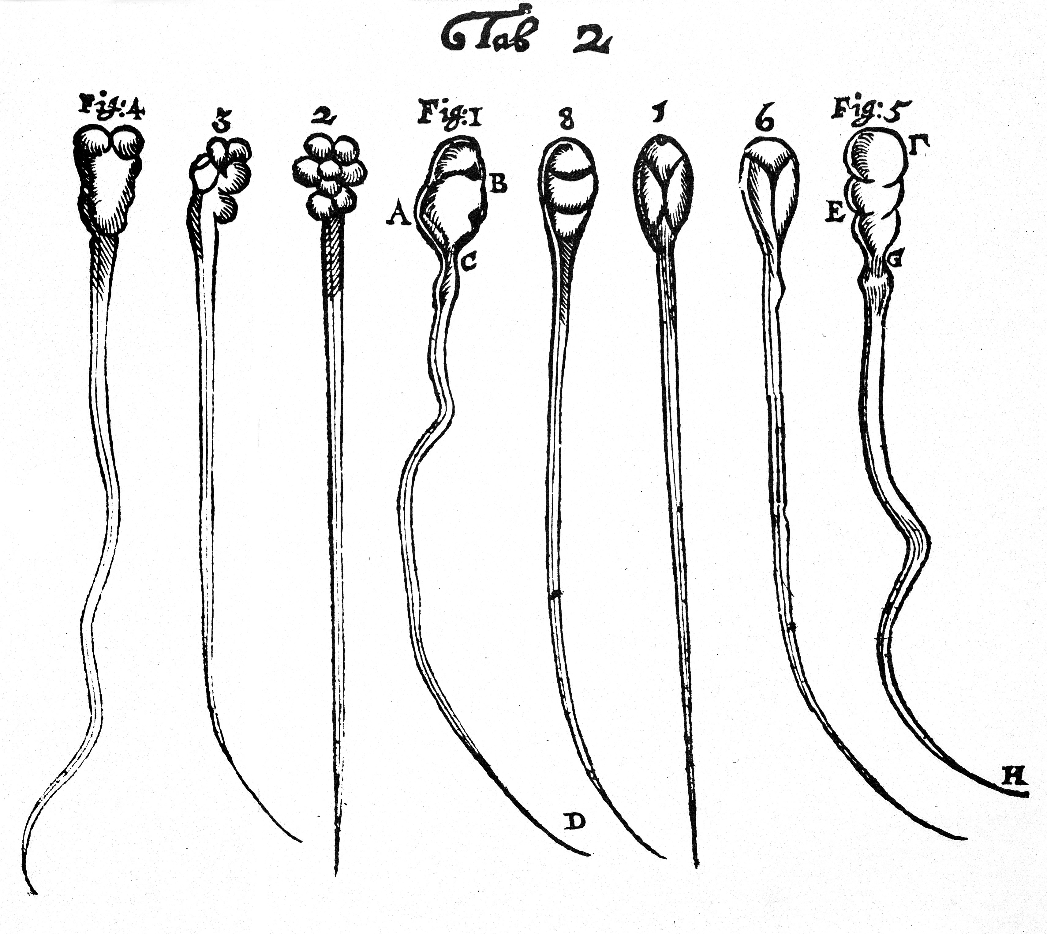
Additionally, in 1677, Leeuwenhoek was also the first person to observe sperm under a microscope when he observed his own ejaculate and noticed tiny wriggling things inside it. These “animalcules”, as he called them, were utterly unknown before this time and, despite contemporary prudence and his own reluctance, Leeuwenhoek shared their discovery with the Royal Society, leading to the first investigation into the field of sperm biology.

Microscopic observations of sperm from Leeuwenhoek.
Seeing is believing
Despite these exciting discoveries, confidence in microscopes remained contentious for nearly 200 years. As mentioned above, the issue here was that these early examples were often blurry and distorted, which made some researchers reluctant to trust them. The situation changed, however, in 1830, when Joseph Jackson Lister, a wine merchant, microscopist, and father of the famous pioneer of antisepsis (also called Joseph Lister), overcame the aberrations that plagued microscopes in collaboration with William Tulley, an instrument manufacturer.
The implications were massive; the scientific community was now armed with a superior device that offered far clearer views of the hidden world. Although it is not easy to tie a neat causal link between this development and subsequent scientific breakthroughs, it nevertheless marked the start of a century of discoveries whereby scientists increasingly came to rely on these instruments in their laboratories and in the field.
During the 1830s, microscopes enabled an in-depth examination of cells and the development of cell theory in medical and biological research. Increased confidence in microscopic imagery meant scientists were able to explore and describe aspects of the body in greater detail. In particular, between 1838 and 1839, Matthias Schleiden and Theodor Schwann, two German scientists, proposed that cells were the fundamental units from which plants and animals were made. Schwann went as far as proposing that an understanding of cellular behavior would change how we understand the body in health and sickness.
His theory was picked up by Rudolf Virchow, an influential pathologist, who championed microscopes as crucial instruments in the scientific toolkit. Virchow progressed cell theory by stating that all cells develop from existing cells and also showed that, when cells go wrong, they can produce diseased tissue.
Microscopes and public health
Microscopes also played a crucial role in the wider context of contemporary public health, albeit at a staggered rate. In the late 1840s and 1850s, cholera epidemics had washed across Britain and many other parts of Europe, killing thousands as it spread. At the time, several doctors conducted microscopic analyses of public water supplies to see what was going on. Some detected a distinct organism that they thought could be responsible for the disease, while others did not. For instance, Frederick Brittan, Joseph Swayne, and William Budd, physicians in Bristol, identified what they believed was a cholera-causing organism in 1849, but it was not supported by others – it is likely they actually identified a fungus.
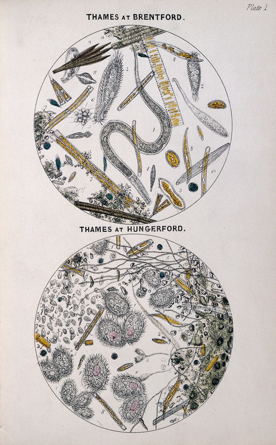
Then, in 1850, the British physician, Arthur Hill Hassall, published his pithily titled book The microscopic examination of the water supplied to the inhabitants of London and the suburban districts, which demonstrated how unclean London water was. Although Hassall’s work informed much discussion at the time and influenced the 1852 Metropolis Water Act, real change took several decades to manifest. In 1884, Robert Koch, the famous German microbiologist, announced the discovery of the causal organism responsible for cholera, but even then, the most significant driving forces behind public reforms were political in nature, rather than scientific.

Microscopic examination of water in London.
Nevertheless, the ability to identify the causal agents responsible for diseases changed the nature of medicine, our understanding of illness, and, eventually, how to treat and prevent them.
Microscopes of the 20th century and beyond
By the 20th century, microscope technologies and the advances in mathematics led to significant changes for viewing the microscopic world. In particular, scientists realized that, if they wanted to enhance the resolution of modern instruments, they would need a different source of light than the type used in traditional microscopes.
An important step in this story came in 1931, with the invention of the first transmission electron microscope (TEM), which was designed and built by Ernst Ruska and Max Knoll. These microscopes rely on electrons rather than light and send electrons through a condenser lens before they reach the specimen being observed, close to what is called the objective lens. TEMs are capable of magnifying objects as small as the diameter of an atom.
Then, in the years between 1939 and 1942, Ruska developed the first scanning electron microscope (SEM), which created images by detecting electrons that are reflected off a specimen (rather than passing through them, as with TEM).
The two different instruments have their own strengths. TEMs are useful for examining the inner structure of a sample, such as crystal structures, morphology, and stress state information, while SEMs provide details of an object’s surface and composition.
Over the next decades, additional advances in visualization techniques led to the introduction of other instruments, such as the first computerized axial tomography (CAT) scanner and the confocal laser scanning microscope. Increasingly, these devices allow us to see more detail within microscopic specimens, but they rely less on traditional lenses as are used in optical microscopes.
However, in the mid-1990s, Stefan Hell, a director at both the Max Planck Institute for Multidisciplinary Sciences in Göttingen and the Max Planck Institute for Medical Research in Heidelberg, Germany, invented the super-resolution microscope. This new optical microscope captures images with significantly higher resolution than anyone had thought possible. In 2014, Hell, Eric Betzig, and W.E. Moerner were awarded the Nobel Prize in Chemistry for developing super-resolution fluorescence microscopy, which brings optical microscopy into the nanodimension (to scales smaller than a billionth of a meter).
These developments have opened up incredible possibilities for our understanding of biological functions at a molecular level, and none of it would have been possible without the long history of lenses that continue to make the invisible visible.
All “explainer” articles are confirmed by fact checkers to be correct at time of publishing. Text, images, and links may be edited, removed, or added to at a later date to keep information current.
Source Link: Microscopic Investigations That Led To Macroscopic Discoveries: How Lenses Changed Science