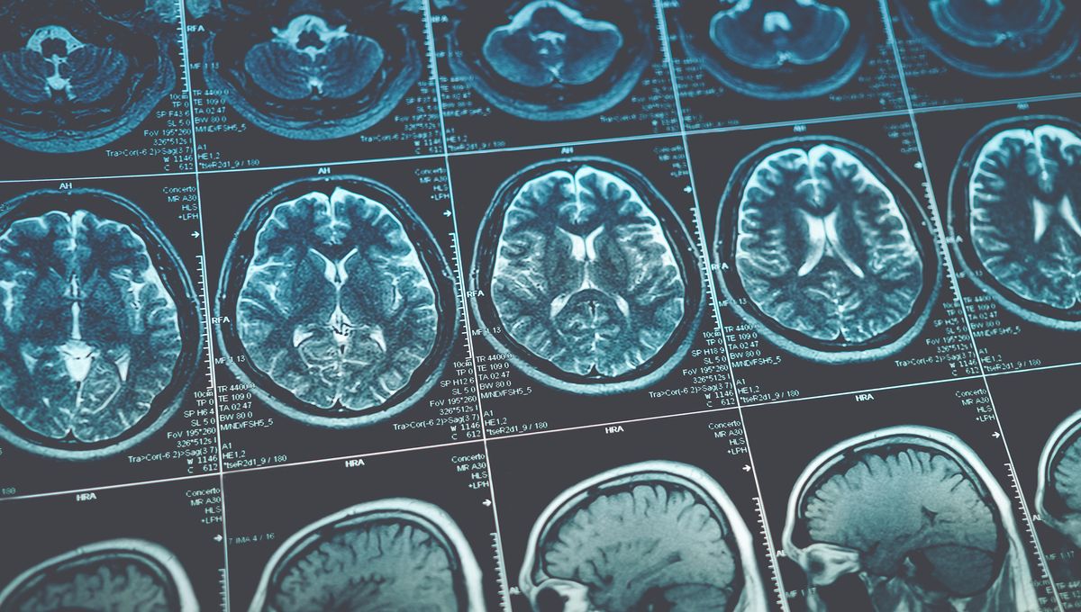
Using a new analytical technique, scientists have been able to study brain images from more than 6,000 children to identify connectivity patterns that are common to people with attention-deficit/hyperactivity disorder (ADHD).
Most of our behaviors are controlled by coordinated communication between neurons in different areas of the brain. Neuroscientists can get a sense of how the regions of the brain orchestrate complex functions by observing neural activity in a resting-state functional magnetic resonance imaging (rs-fMRI) scan.
“Resting state” means exactly what it sounds like – these scans are carried out while the subject is at rest, not being asked to perform a particular cognitive task or think any particular thoughts. Assuming you’re not claustrophobic, and don’t mind keeping perfectly still, it can be a fairly pleasurable experience.
The data derived from rs-fMRI scans is invaluable to scientists studying a whole range of neurological disorders and conditions. By comparing scans from individuals with conditions like ADHD, for example, with those of neurotypical people, it’s hoped we’ll be able to identify patterns that can explain some of the features of these conditions.
However, this type of research into ADHD has so far been hindered by small sample sizes and inconsistent methods, so it’s been difficult to draw any firm conclusions. A recent study led by Michael Mooney at Oregon Health & Science University sought to change all that.
Using several large-scale datasets, the team developed a new way of analyzing imaging data covering broader areas of the brain than ever before. They called this a polyneuro score (PNRS).
“Our findings demonstrate a robust association between brain-wide connectivity patterns (PNRS) and 554 ADHD symptoms in two independent cohorts,” they explain in their paper.
The authors go on to explain how their approach could be used to glean better insights from even small datasets, and could also be used to identify mechanisms that may be shared across different neurological and psychiatric conditions – for example, could it be the case that an ADHD-typical PNRS is predictive of depression symptoms? This could help identify patients at risk of comorbidities.
ADHD diagnoses are on the up and we’re learning more about the condition every day, but there are still some significant gaps in our knowledge about the underlying neurobiology. Collecting lots of imaging data is only one piece of the puzzle – you also need ways to use that data that answer the questions you have. The authors of this study hope their methods will make that more achievable, for ADHD and many other conditions.
The study is published in The Journal of Neuroscience.
Source Link: Over 6,000 Scans Reveal What ADHD Looks Like In The Brain