The winners of Nikon’s Small World Photomicrography Competition 2022 have just been announced, showcasing the most beautiful images of the tiny world that exists under a microscope.
This year’s top prize was given to Grigorii Timin, supervised by Dr Michel Milinkovitch at the University of Geneva, for his stunning detailed image of a hand of an embryonic Madagascar giant day gecko.
With its vibrant colors and simple composition, the image is also evidence of technical prowess, requiring high-resolution microscopy and image-stitching to capture. The result shows just how complex these tiny structures are, with the hand’s bones, tendons, ligaments, and skin shown in cyan and its blood cells highlighted in the orangey colors.
“This embryonic hand is about 3 mm (0.12 in) in length, which is a huge sample for high-resolution microscopy,” said Timin in a statement. “The scan consists of 300 tiles, each containing about 250 optical sections, resulting in more than two days of acquisition and approximately 200 GB of data.”
“This particular image is beautiful and informative, as an overview and also when you magnify it in a certain region, shedding light on how the structures are organized on acellular level,” he explained.
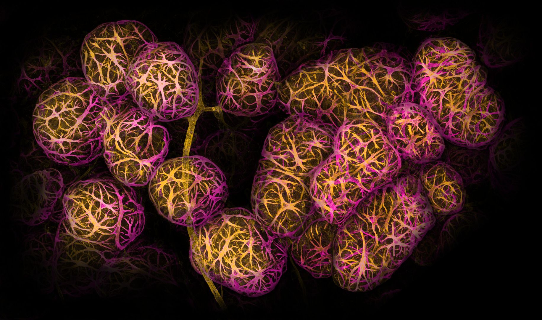
2nd Breast tissue showing contractile myoepithelial cells wrapped around milk-producing alveoli. Image credit: Dr Caleb Dawson/Nikon Small World Photomicrography Competition 2022
This year marks the 48th annual Small World Photomicrography Competition which saw almost 1,300 entries from 72 countries. Four judges went through all of these submissions and evaluated them based on originality, informational content, technical proficiency, and visual impact.
“At the intersection of art and science, this year’s competition highlights stunning imagery from scientists, artists, and photomicrographers of all experience levels and backgrounds from across the globe,” added Eric Flem, Communications and CRM Manager, Nikon Instruments.
Second place was awarded to Dr Caleb Dawson for an image of breast tissue showing contractile myoepithelial cells wrapped around milk-producing alveoli, while third place was swept up by Satu Paavonsalo and Dr Sinem Karaman for their image of blood vessel networks in the guts of a mouse
You can see some of the top 10 images below, plus dozens of other winners and honorable mentions here. Also, be sure to check out this year’s Nikon Small World in Motion competition right here.
Autofluorescence of a single coral polyp (approx. 1 mm). Image credit: Brett M. Lewis/Nikon Small World Photomicrography Competition 2022
A fly under the chin of a tiger beetle. Image credit: Murat Öztürk/Nikon Small World Photomicrography Competition 2022
Unburned particles of carbon released when the hydrocarbon chain of candle wax breaks down. Image credit: Ole Bielfeldt/Nikon Small World Photomicrography Competition 2022
Slime mold (Lamproderma). Image credit: Alison Pollack/Nikon Small World Photomicrography Competition 2022
Long-bodied cellar/daddy long-legs spider (Pholcus phalangioides). Image credit: Dr Andrew Posselt/Nikon Small World Photomicrography Competition 2022
3rd place: Blood vessel networks in the intestine of an adult mouse. Image credit: Satu Paavonsalo & Dr Sinem Karaman/Nikon Small World Photomicrography Competition 2022
Source Link: The Mind-Blowing Winners Of Nikon's Small World Photo Competition 2022
